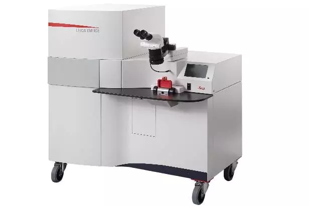
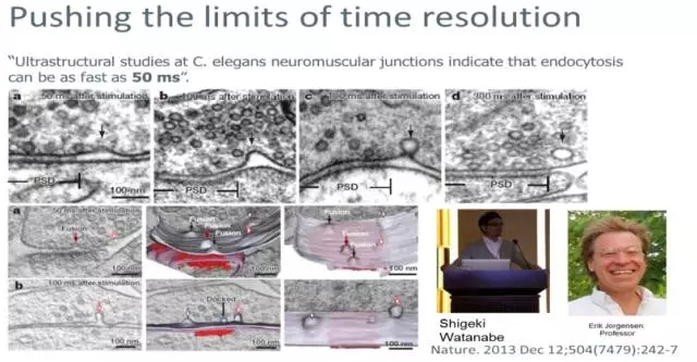
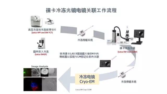
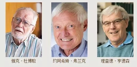
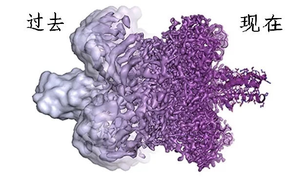
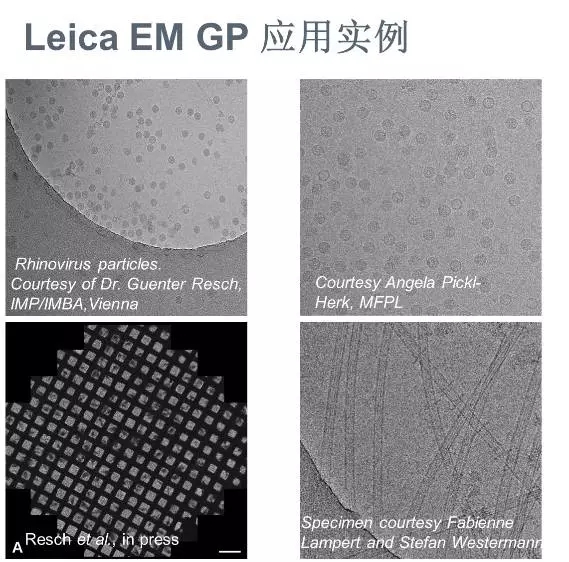
2017 Nobel Prize in Chemistry The 2017 Nobel Prize in Chemistry was awarded to Richard Henderson, Joachim Frank and Jacques Dubochet for their development in cryo-electron microscopy. The outstanding contribution made. Resolution comparison They simplified the cryo-electron microscopy technique and applied it to the direction of biomolecular imaging, breaking the long-standing analytical protein that relied on traditional X-ray crystallography (NMR) and nuclear magnetic resonance (NMR), which made us very It is possible to obtain detailed images of life complex mechanisms at atomic level resolution. (Source: Martin Hogbom) Since frozen sample preparation is so important, is there an instrument that can improve the repeatability and success rate of our experiments? Have! That is our Leica's carrier network into the freezer GP. The Leica EM GP is designed for cryo-electron microscopy sample preparation for the preparation of vitrified liquid samples. Sample suspensions such as viral particles, proteins and other cellular components. Where is the future of cryo-electron microscopy technology? In addition to biological macromolecules such as proteins, biological samples also have important aspects of cells and tissues. Even though many important protein structures are currently analyzed by Amy's level, they are all purified from the original position, just like a leaf leaves the big tree. The research is deep, and it is only a leaf cover. Don't say that the position of the leaves in the forest is difficult to say, even on the growth site and structure of which particular tree. Therefore, the analysis of large-scale high-resolution fine structures such as cells or tissues has broader biological significance. So what is the progress of our research on their sample preparation methods? This again has to mention Professor Dubocher of this Nobel Prize. He not only established a single-particle frozen sample preparation technology, but also made outstanding contributions at the cell organization level, especially the development and gradual improvement of CEMOVIS (Cryo-Electron Microscopy Of VItreous Sections), achieving cell tissue in a near-physiological state. In situ observation. CEMOVIS is not chemically fixed, has no dyeing, and has no accumulation of biological components; it maintains the in situ, moisture-fixed glassy state of the organism. The SEM image obtained with CEMOVIS looks very different from the conventional sample image: the structure is smoother and more homogeneous, and the details are more obvious. So what is its technical route? High pressure freezing + frozen section + cryo-electron microscope. Professor Du Boche's initial work was performed using Leica's EM PACT (Leica's first generation of high-pressure cryostat) to freeze the samples, cryosectioning with FCS Ultracut S cryomicrotome, and finally observing under TEM. (See below). As a leader in the field of high pressure refrigeration, Leica has developed to the third generation of Innovative Cryo-fixation Equipment. The high-pressure freezer is cooled by liquid nitrogen, and the pressure source is provided by the air compressor. The physical pressure can be used to achieve a partial pressure of 2100, thereby achieving high-pressure freezing and fixing of the aqueous sample. After the freezing and fixing, the sample is subjected to subsequent treatment (freezing replacement/normal temperature). Ultrathin sections, frozen ultrathin sections or frozen fractures, etc.) are observed by electron microscopy. The high-pressure freezer can preserve the original real information of the living state of the sample in the millisecond level, the protein structure, enzyme/antigen activity is unchanged, the soluble ions and small molecules are effectively fixed, etc., which effectively avoids the conventional chemical fixation. Illusion. In addition to the work of CEMOVIS in combination with cryosectioning, it can also be used to observe the internal ultrastructure of the sample or perform immunological work in combination with cryo-fracture, cryopreservation, etc. In addition, it can combine light and electrical stimulation to open a new door for neurophysiologists. Now, the frozen sample is gradually deep into the hearts of the people. After the frozen sample is processed, can it be explored at the same time under the light microscope? Leica EM Cryo CLEM (Cryo Correlative Light Electron Microscopy) freeze-mirror electron microscope combined system to maintain the correlation between light and electron image in the original state of the sample, and to collect fluorescence microscopic images and high-resolution electron microscope images at the same sample position. One. Its main technical route is: Prepare frozen ultrathin sections (or freeze samples by input freezing technique) by freezing sample preparation technology (high pressure freezing, preserving the original subcellular structure information of the sample), and then observe the fluorescence by means of frozen light microscope The position of the mark is recorded, the position of interest is recorded, and then the in-situ electron microscope observation is performed in the frozen TEM, thereby realizing the technique of combining light and electron microscope. Remarks: High-pressure freezing and freezing ultra-thin sections for freezing fixed tissue samples, such as tissue samples infected with virus. The frozen EM GP is used to freeze single-particle viral macromolecules, or protein macromolecular particles. At the moment, whether you are doing a single particle or a cell organization, do you want to try our Leica GP, ICE and Cryo CLEM? Call us at 400-630-5902! To the true picture, the sample is first! May our Leica Microsystems wish you the best! About Leica Microsystems Leica Microsystems Leica Microsystems is a global leader in microscopy and analytical science instruments and is headquartered in Wetzlar, Germany. It mainly provides professional scientific instruments such as research-grade microscopes in the field of microstructure and nanostructure analysis. Since the establishment of the company in the 19th century, Leica has been widely recognized by the industry for its quest for optical imaging and its innovative spirit of continuous improvement. Leica is a global leader in multiple microscopy in composite microscopes, stereo microscopes, digital microscopy systems, laser confocal scanning microscopy systems, electron microscopy sample preparation and medical surgical microscopy. Leica Microsystems has seven product development and production sites around the world and has service support centers in more than 20 countries. Leica has regional branches or sales branches in more than 100 countries around the world, and has established a comprehensive dealer service network system throughout the world. Lab Researching Microscope ,Microscope Lab,Biological Microscope,Laboratory Microscope Ningbo ProWay Optics & Electronics Co., Ltd. , https://www.proway-microtech.com
Among them, it is worth mentioning that Professor Du Boche's work: He drops a small amount of protein solution onto the copper grid, and quickly inserts it into liquid ethane cooled with liquid nitrogen, so that the protein particles are rapidly cooled by the glassy ice. The packaged, to the greatest extent, preserved the true original state of the protein particles, and obtained high-quality samples of cryo-electron microscopy, which greatly promoted the promotion of cryo-electron microscopy. 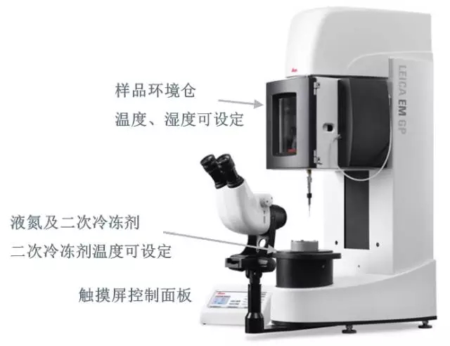
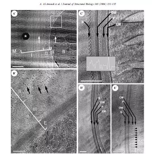
Celluar CryoEM